

Synchrotron-based Research on Geomaterials
The availability of synchrotron radiation for research in geoscience opens new experimental possibilities by overcoming the former limitations such as detection limit and spatial resolution. Our group uses a broad energetic range of synchrotron radiation from high-energetic X-rays down to low-energetic infrared radiation for research mostly in-situ at high pressures in diamond anvil cells (DAC).
X-ray diffraction
The very high brilliance X-rays available at the modern synchrotron sources, such as PETRA III at DESY in Hamburg, allows us to determine (by measuring X-ray diffraction, XRD) the compression behavior of geomaterials compressed in the diamond anvil cell (DAC) up to very high pressures relevant for the deep interior of the Earth. We are currently performing studies of the pressure-volume (P-V) equation of state of hercynite (FeAlO4) the iron end-member of the upper mantle peridotitic spinel and of the 3.65 Å phase, MgSi(OH)6 the product of decomposition of the 10 Å phase and a relevant candidate for the deep transport of hydrogen in the mantle through subduction. Moreover we examine the synthesis and properties of new materials containing Mg-Fe-N and Si-Ge.
Contact
Dr. Sergio Speziale
Dr. Hans Josef Reichmann
X-ray fluorescence
X-ray fluorescence (XRF) is the emission of characteristic "secondary" (or fluorescent) X-rays from material that has been excited by exposure to high-energy X-rays. The phenomenon is widely used for elemental and chemical analysis. Due to the high penetration depth of hard X-rays in matter, XRF can be used to determine element concentrations in complex sample environments, such as high-pressure cells. The high brilliance of synchrotron radiation strongly improves sensitivity and spatial resolution of XRF analyses and allows sampling of trace elements in materials that cannot be quenched to ambient conditions, such as aqueous fluids.
Contact
Dr. Christian Schmidt
Partner
Prof. Dr. Max Wilke, Universität Potsdam
X-ray absorption spectroscopy (XANES, EXAFS)
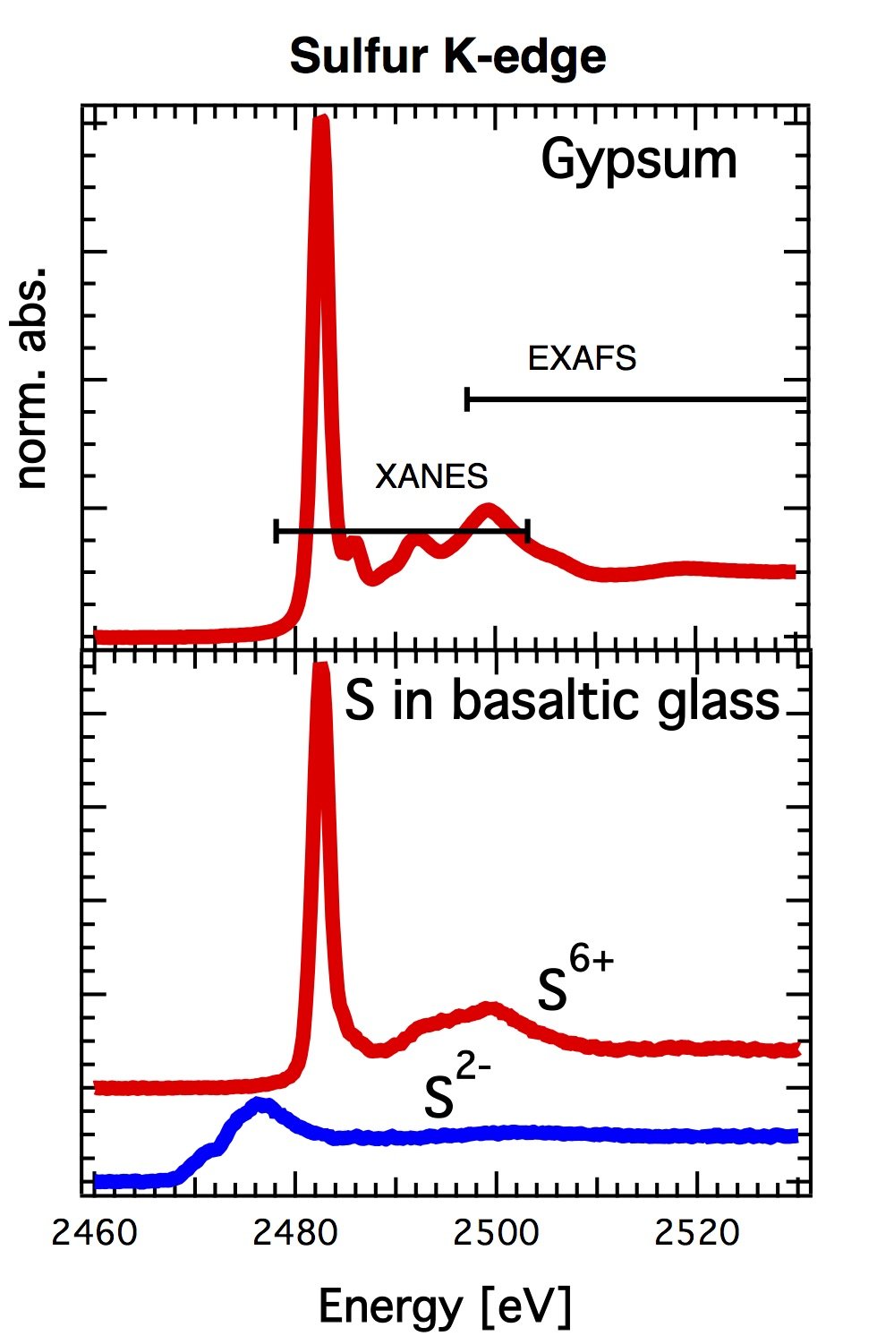
X-ray absorption spectroscopy uses x-rays to probe the physical and chemical structure of matter on the atomic scale. X-ray absorption spectroscopy is element-specific, which means that X-rays are chosen to be at and above the binding energy of a particular core electronic level of a specific element. Because most elements have core-level binding energies in the X-ray regime, nearly all elements except the lightest can be studied with this technique. The fine structure observed in the X-ray absorption is due to interaction of the photo-electron with unoccupied electronic states of the absorbing element or due to backscattering from neighboring atoms. The fine structure is usually divided into two regions: XANES, X-ray Absorption Near Edge Structure: Fine structure observed close to the binding energy, which is assigned to excitation of the photo-electron to localized states or to multiple backscattering of the photo-electron by neighboring atoms. The XANES region is sensitive to oxidation state, coordination and site geometry of the absorbing element. EXAFS, Extended X-ray Absorption Fine Structure: Fine structure observed at energies far above the binding energy, which is assigned to single backscattering of the photoelectron by neighboring atoms. Analysis allows determination of number of neighbors and distances to neighbors of the absorbing element in crystalline and non-crystalline samples. Both, XANES and EXAFS, are applicable to concentrated and dilute samples. Due to the high penetration depth of hard X-rays, measurements can be performed even at in-situ conditions using complex sample environments, such as pressure cells or reaction cells, at least for absorption edges above ca. 6 keV.
X-ray Raman scattering (XRS)
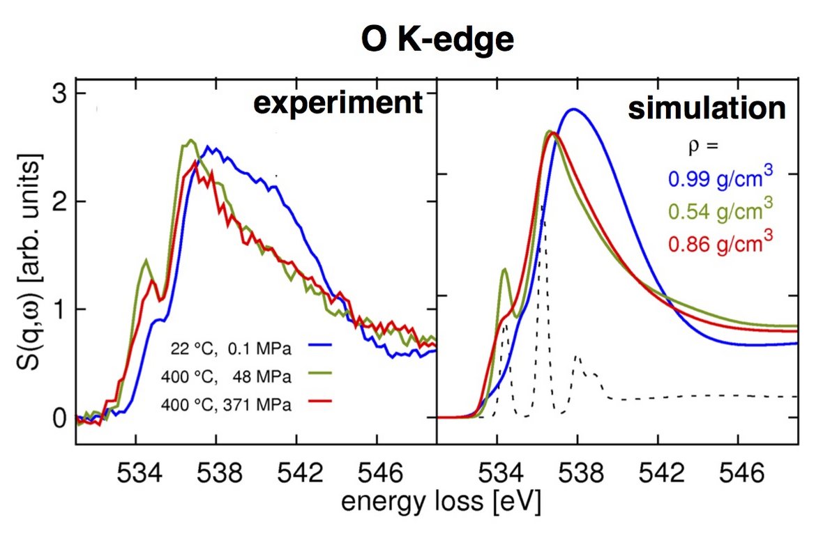
X-ray Raman scattering (XRS) is an inelastic x-ray scattering process, in which a high-energy X-ray photon transfers energy to a core electron, exciting it to an unoccupied state. The process is in principle analogous to X-ray absorption, but the energy transfer plays the role of the X-ray photon energy absorbed in X-ray absorption. Because the energy of the probing X-ray can be chosen in the hard X-ray regime, certain constraints of soft X-rays are overcome. For example, soft X-ray studies usually require vacuum environment. This makes X-ray absorption studies of samples that are not stable in vacuum, such as many liquids, impossible. One of the most exciting applications of X-ray Raman scattering is the study of soft X-ray absorption edges at high pressure. The employed high-energy X-rays easily pass through a high-pressure apparatus like a diamond anvil cell and reach the sample inside the cell.
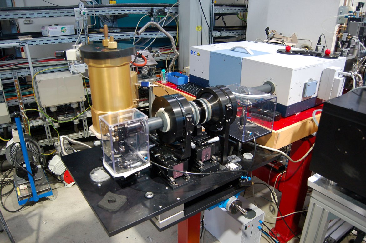
Infrared spectroscopy: Measurements in Diamond Anvil Cells (DACs)
We work at the Synchrotron Infrared (SR-IR) Beamline of the Helmholtz Centre Berlin, Bessy II, to perform:
Synchrotron-based absorption measurements in the far-infrared (SR-FIR) spectral range on materials in DACs
To investigate pressure-induced phase transitions diamond anvil experiments are widely used in combination with X-ray diffraction but can also be performed using FTIR spectroscopy. Infrared spectroscopy is a very sensitive probe for information on the local microscopic structure and bonding in minerals. Combined with a high brightness synchrotron source infrared micro-spectroscopy offers the opportunity to achieve the spatial resolution approaching the diffraction limit. We apply Synchrotron-based THz/FIR spectroscopy to investigate pressure-induced phase transitions of geomaterials in diamond-anvil cells (DAC) up to 30 GPa at Bessy II. To do so, we developed in cooperation with Dr. U. Schade from the Helmholtz Centre Berlin, Bessy II, a THz/FIR microscope adapted to a Bruker Vertex 80v spectrometer, which enables us to perform FIR measurements under vacuum. Experiments on wadsleyite, Sr-anorthite, leucite, ilvaite and various carbonates were successfully performed and showed that this method is very powerful.
Synchrotron-based absorption measurements in the mid-infrared (SR-MIR) spectral range on oriented single crystal at ambient conditions and in DACs
A powerful tool for detecting traces of hydrogen in nominally anhydrous minerals (NAMs) is IR spectroscopy. However, IR-spectroscopy is often hampered by the relative small sample size (20-30 μm) and the comparatively large beam size needed to get an acceptable signal to noise ratio (about 60 μm). With synchrotron radiation in combination with micro-spectroscopy (Nicolet Continuum Microscope) we can achieve a spatial resolution down to 5 x 5 µm and, e.g. the OH-distribution in crystals or the high-pressure behavior of geomaterial can be studied on a much smaller scale and to much higher pressures, respectively, than usually applied using conventional IR radiation.
Contacts
Prof. Monika Koch-Müller
Dr. Ilias Efthymiopoulos
Partner
Dr. Ulrich Schade, Helmholtz Zentrum Berlin, Institut Methoden der Materialentwicklung